In the first procedure of its kind, a team from Brigham and Women’s Hospital and Boston Children’s Hospital successfully performed an in utero cerebrovascular surgery to treat a vascular malformation in an infant’s brain before birth.
A recent Brigham press release and a paper published in Stroke described the successful procedure, which took place in an obstetric operating room with a maternal-fetal medicine specialist and fetal radiologist. Louise Wilkins-Haug, MD, PhD, director of the Division of Maternal-Fetal Medicine at the Brigham, and Carol Benson, MD, staff radiologist and former co-director of the Brigham’s High-Risk Obstetrical Ultrasound Service, and Darren Orbach, MD, PhD, chief of Neurointerventional Radiology at Boston Children’s Hospital and principal investigator for an ongoing clinical trial on the intervention, were co-authors.
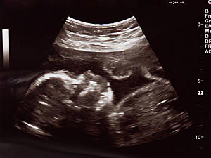
Vein of Galen Malformation
The partnership between Boston Children’s Hospital and the Brigham’s Fetal Therapy Program arose as part of an ongoing clinical trial to treat vein of Galen malformation (VOGM) in utero.
VOGM is a rare and potentially deadly developmental condition that occurs when misshapen arteries in the brain connect directly to veins instead of capillaries, causing high-pressure blood to flow into the veins. This can prevent the infant’s brain from adequately draining and lead to brain injury or severe loss of tissue in the brain.
VOGM is typically treated after the infant is born, but in many cases, brain damage has already occurred, and many infants die of heart failure within the first few days of birth. Thus, there is an effort to correct the malfunction before birth to prevent brain injury.
About the Procedure
A multidisciplinary team from radiology, neurointerventional radiology, anesthesiology, and maternal-fetal care performed the in utero embolization on the fetus with VOGM at 34 weeks and two days gestational age. As part of an ongoing clinical trial, the procedure was performed with U.S. Food and Drug Administration oversight.
The team used ultrasound-guided transuterine embolization to address the infant’s VOGM before birth. The procedure was successful, and the newborn did not require additional surgery after birth. Brain MRI revealed no strokes, fluid buildup, or hemorrhage, often occurring in VOGM. After several weeks in the NICU, the infant was discharged and is hitting all their milestones.
This proof of concept procedure could mark a paradigm shift in the management of VOGM, where the malformation is repaired before birth to reduce the risk of long-term brain damage or death.