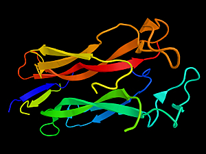
T helper cells that produce interleukin-17 (Th17 cells) are an important subtype of CD4+ T cells in the intestine. Th17 cells enhance the gut epithelial barrier function, protecting the host from bacterial and fungal infections. On the other hand, they can be pathogenic in chronic inflammatory diseases such as multiple sclerosis, rheumatoid arthritis, and inflammatory bowel disorder (IBD).
Brigham and Women’s Hospital recently determined how epithelial cells promote Th17 cell generation in response to bacterial colonization as a step toward better understanding the treatment—and even prevention—of these diseases.
Richard S. Blumberg, MD, vice chair for Research in the Department of Medicine at the Brigham, Jinzhi Duan, PhD, an instructor in the Department of Medicine at the Brigham, and Juan D. Matute, MD, instructor in the Department of Pediatrics at Massachusetts General Hospital, and colleagues report in Immunity.
Background
Intestinal epithelial cells (IECs) form a protective interface between intestinal microbes and leukocytes. However, they’re secretory and susceptible to endoplasmic reticulum (ER) stress. Dr. Blumberg and colleagues previously demonstrated in Nature that the unfolded protein response (UPR) enables highly secretory epithelial cells to maintain homeostasis.
Conversely, in that study and others, defective UPR in IECs promoted inflammatory ER stress that led to IBD.
Using several mouse models, the research team showed in the new study that ER stress and the associated UPR in IECs underlies microbially induced Th17 development.
Detail of the Findings
Key observations from the mouse studies were:
- Microbe adhesion to IECs drove ER stress that caused a UPR and induced Th17 cells in the intestine; these cells were associated with non-pathogenic gene expression signatures in germ-free mice and germ-reduced (antibiotic-treated) mice
- UPR activation in IECs increased their production of both reactive oxygen species (ROS) and purine metabolites
- Intriguingly, the most potent purine metabolite was xanthine, a mild stimulant in coffee, tea, and chocolate
- Treating mice to reduce either ROS or xanthine production decreased the number of Th17 cells associated with a UPR
The team confirmed a positive relationship between ER stress signatures and Th17 cell-related gene expression using published RNA sequencing data from adults and children with IBD.
Commentary
Intriguingly, IEC-ER stress-induced Th17 cells didn’t have pathogenic signatures in mice with few or no microbes. Even though IEC-related ER stress is commonly observed in IBD, those cells might have a homeostatic role.
In moving toward the clinical translation of the results, it will be interesting to see whether IBD-associated Th17 cells overlap with the IEC-ER stress-induced Th17 cells found in these mouse models. More generally, the findings may lead to better understanding of gut health and give insight into the pathogenesis of inflammatory diseases.
