Loss of smell is a cardinal finding in several prevalent inflammatory and infectious diseases, including COVID-19. Clinical studies of olfaction typically rely on subjective tests, but some scents are not familiar to individuals of all cultures, and the tests are susceptible to patient response bias.
In a proof-of-concept study, Stella E. Lee, MD, director of the Sinus Center in the Division of Otolaryngology–Head & Neck Surgery at Brigham and Women’s Hospital, and colleagues demonstrated a safe, portable, objective method for measuring smell function and its relationship with cognitive processing. The findings appear in Frontiers in Allergy.
Background
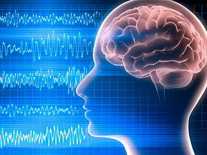
Electroencephalography (EEG) is the usual objective method for measuring the sense of smell. It records electrical signals that reflect the brain’s response to olfactory stimulation. That spatial information can distinguish between normosmic, hyposmic, and anosmic individuals.
EEG can be supplemented with magnetoencephalography (MEG), which primarily detects magnetic fields induced by intracellular currents. Unlike EEG, MEG provides temporal information, improving the estimates of what brain regions respond to scents. MEG data are often superimposed on MRI scans to provide a user-friendly anatomic interface for interpretation.
Dr. Lee’s team built a portable olfactometer that combines EEG and MEG/MRI. The pilot study showed it could identify odorant-specific “cortical signatures” during olfactory stimulation that can be visualized with spectral analysis.
Methods
The study involved three healthy subjects (two males and one female; mean age 31) who were normosmic according to the University of Pennsylvania Smell Identification Test.
The novel olfactory stimulator delivered two synthetic odorants into a CPAP mask: 2% phenethyl alcohol (rose) and 0.5% amyl acetate (banana). It was programmed to present each odorant over increasing increments, followed by rest periods. Cortical activity was measured with MEG. High-resolution MRI images were obtained for each subject and were co-registered with the MEG data.
Spectral analysis was used to calculate the oscillatory power in different bands (alpha, beta, theta, delta, and gamma) separately for each olfactory condition and for rest.
Results
In the initial subject, the largest effect during olfactory stimulation was found in the alpha band. The results here are reported for that band relative to the rest:
- Olfactory cortex: Approximate 40% increase in power bilaterally during both phenethyl alcohol and amyl acetate stimulation
- Somatosensory cortex: Approximate 30% decrease in power, right greater than left, with both odorants
- Left orbitofrontal cortex: Approximate 25%–30% increase in power with phenethyl alcohol but not amyl acetate
- Left medial frontal cortex: 5%–10% greater increases in power with phenethyl alcohol than amyl acetate
- Left precentral gyrus: 25%–30% greater increases in power with phenethyl alcohol than amyl acetate
Potential Applications
Reliable, unique olfactory signatures of odorants may be centrally observed with this new tool and be useful for the following:
- Improving the diagnosis and management of chronic rhinosinusitis, COVID-19, and other hyposmia and anosmia disorders that have infectious or inflammatory causes
- Managing certain neurodegenerative diseases where the sense of smell is affected, such as using the loss of smell to screen for preclinical Alzheimer’s disease and identify which patients with Parkinson’s disease are likely to have a rapid neurologic decline
- Studying olfactory dysfunction in general, including the connection between smell and cortical function
