Cerebral proliferative angiopathy (CPA) is a cerebral vascular malformation with distinctive features: large size, absence of dominant feeders or flow-related aneurysms, transdural supply of healthy and pathological tissues, brain parenchyma intermingled between vascular spaces, capillary angioectasisa, and only moderately enlarged veins.
CPA usually presents with ischemia, but when hemorrhage occurs, there is a higher risk of re-hemorrhage.
Jakob V. E. Gerstl, MBBS, a research fellow in the Department of Neurosurgery at Brigham and Women’s Hospital, Rose Du, MD, PhD, director of the Cerebrovascular Surgery Program in the Department, and colleagues recently presented in The Neurohospitalist one of the few reports of long-term follow-up of a patient with hemorrhage in CPA: 32 years of data on a patient who had recurrent hemorrhage.
Initial Presentation
A 19-year-old male presented to an outside institution with focal awareness seizures characterized by right hand twitching. Work-up revealed a large left hemispheric vascular lesion, which was diagnosed as an arteriovenous malformation. The patient was managed conservatively with antiepileptics.
First Hemorrhage
At age 28, the patient woke at night with a severe headache, nausea and vomiting. Noncontrast head CT revealed an intraventricular hemorrhage. The patient was admitted to the Brigham neurosurgical unit for observation, where he remained stable without the need for an external ventricular drain (EVD).
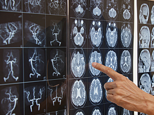
MRI without contrast confirmed a 9-cm vascular lesion in the left hemisphere. It was deemed inoperable and the patient was discharged home nine days after admission. Later he developed weakness and contracture in the right hand, and he continued to have intermittent focal awareness seizures characterized by right hand shaking.
Second Hemorrhage
At age 43, the patient presented again following an episode of nausea and vomiting. Head CT confirmed re-hemorrhage into the ventricles and development of ventriculomegaly. The patient was admitted for monitoring but still did not require an EVD.
MRI with contrast re-demonstrated a 9-cm left hemispheric vascular lesion and this time showed brain parenchyma intermingled between vascular spaces. Findings of MR and conventional angiography were consistent with CPA:
- Abnormal, ectatic feeders from the left anterior cerebral artery, left internal carotid artery and the left middle and left posterior cerebral arteries
- Multiple superficial draining veins to the superior sagittal sinus along with deep venous drainage to a large ectactic vein that drained to the internal cerebral vein
- Absence of a dominant feeder
- A large hemispheric nidus
- Relatively small draining veins
The patient was stabilized and discharged on hospital day 4. However, he developed progressive hemiparesis and aphasia. During the same period, fluid attenuated inversion recovery (FLAIR) MRI showed increasing perilesional hyperintensities.
Third Hemorrhage
At age 51, following a seizure and vomiting, the patient presented again. Head CT demonstrated a left frontotemporal intraparenchymal hemorrhage extending throughout the ventricles, more extensive than the second bleed. CT angiography and conventional angiography confirmed the previous CPA findings without any major changes. The FLAIR signal abnormality was further increased on MRI.
The patient was admitted for placement of an EVD. He was otherwise conservatively managed as no clear bleeding point could be identified. The EVD was removed on hospital day 23 and the patient was discharged on day 38 with hemiparesis and aphasia trending toward baseline. He was considered to have a fair prognosis of recovering baseline functional mobility and communication.
Literature Review
A systematic search of PubMed in May 2023 revealed reports on 31 other patients with confirmed hemorrhage in CPA. Two died immediately, three required immediate decompressive craniectomy, and four had immediate endovascular embolization of one or more associated aneurysms.
Most of the other patients were managed conservatively in both the immediate setting of the hemorrhage and at follow-up, but tailored interventions can be considered on a case-by-case basis.