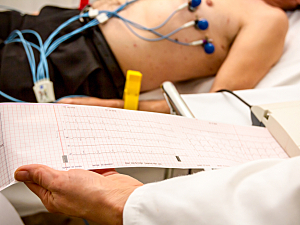
Atrial septal defect (ASD) usually isn’t identified in adults until overt symptoms develop. Echocardiography is highly sensitive and accurate for detection but isn’t feasible for large asymptomatic populations. Electrocardiography (ECG) can be used for screening, but its sensitivity and specificity are currently limited, so a large proportion of patients with ASD are missed.
Shinichi Goto, MD, PhD, an instructor in the Division of Cardiovascular Medicine at Brigham and Women’s Hospital, Yoshinori Katsumata, MD, PhD, of Keio University School of Medicine, and colleagues have developed a deep learning–based model that detects subtle changes in standard 12-lead ECG data to flag patients who should undergo echocardiography for evaluation of possible ASD. In eClinicalMedicine, they report excellent generalizability of the model over three healthcare systems in Japan and the U.S.
Methods
The researchers collected 671,021 ECGs from 80,947 adults who underwent both ECG and transthoracic echocardiography at:
- Keio University Hospital (KUH), a tertiary hospital in an urban area in Japan that is a high-volume center for ASD treatment (n=30,926 patients; 2.24% with ASD; July 2011 to December 2020)
- Brigham and Women’s Hospital (BWH) (n=24,843; 0.29% with ASD; January 2015 to December 2020)
- Dokkyo Medical University Saitama Medical Center (SMC), a primary/secondary care center in a rural area in Japan (n=25,178; 0.27% with ASD; January 2010 to December 2021)
The data from KUH and BWH were used to develop and validate the deep learning–based algorithm to detect ASD, and the algorithm was externally validated using the data from SMC. The algorithm was constructed using convolutional neural networks, a type of artificial intelligence that is trained on thousands of visual patterns and learns to interpret new patterns.
Algorithm Performance
The algorithm showed an excellent ability to distinguish patients with ASD:
- KUH dataset—Area under the receiver operating characteristic curve (AUROC), 0.90
- BWH dataset—AUROC, 0.88
- SMC dataset—AUROC, 0.85 (P=0.52 vs. KUH and P=0.52 vs. BWH)
Other key findings were that:
- The algorithm performed better than predictions based on overt ECG abnormalities (right bundle branch block, right atrial dilation, and any ECG abnormality)
- In all three cohorts, the algorithm’s performance was consistent across age, sex, body mass index, presence or absence of atrial fibrillation, and presence or absence of any ECG abnormalities
- The performance of the algorithm was consistent across races at BWH
Deployment Simulation
The algorithm’s performance was relatively consistent in the three cohorts across various cutoff points. At the cutoff that achieved a 10% positive predictive value (PPV) at KUH, the results were:
- KUH—83% sensitivity and 83% specificity
- BWH—69% and 88%
- SMC—68% and 91%
When the cutoff was adjusted to achieve a 25% PPV at KUH:
- KUH—69% sensitivity and 95% specificity
- BWH—44% and 97%
- SMC—51% and 97%
The algorithm offered great improvement in sensitivity compared with overt ECG abnormalities. For example, in the KUH dataset, the presence of any ECG abnormality had a sensitivity for ASD of 81% and a specificity of 34%; with the algorithm, the sensitivity was 94% at the same specificity.
These results suggest the algorithm will be useful without the need to re-calculate the cutoff value at new institutions. Deep learning–enhanced ECG might someday prove capable of detecting other congenital heart diseases.
