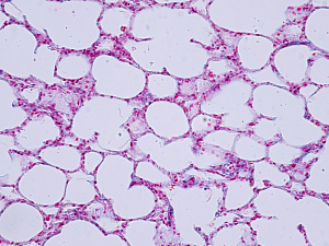
How the pleural epithelium adapts to mechanical stress is relevant to understanding both normal lung function and parenchymal lung disease. However, differences in mechanical stress are difficult to assess because of the nonlinear stress–strain properties of lung parenchyma and large displacements with ventilation. What’s more, no reliable method is known for isolating the pleural epithelium for structural studies.
Now, researchers at Brigham and Women’s Hospital have developed a method for studying the topography of pleural epithelial structure enabled by en face harvest and machine learning. They then used the geometry of pleural epithelial structure to map regional differences in mechanical stress.
Steven J. Mentzer, MD, principal of The Mentzer Lab in the Division of Thoracic Surgery, Betty S. Liu, MD, a fellow in the Lab, and colleagues describe their approach and findings in the Journal of Cellular Physiology.
Methods
The research team’s approach had four main steps:
- In situ silver‐staining of the lung surface minimized processing artifacts, highlighted cell borders, and facilitated imaging with light microscopy
- Pectin, a bioadhesive, was applied to the pleural surface, allowing tissue subjacent to the pleural epithelium to be peeled away in large continuous sheets; this harvest method preserved the spatial distribution and the structure of pleural cells
- Traditional watershed algorithms, which identify gray-level infection to define cell boundaries, were used to study the pleural epithelial images
- A transfer learning neural network was trained to identify cell number, cell area, cell perimeter, and shape in normal adult pleural epithelium—showing agreement with watershed algorithms—and permitted rapid analysis of detailed morphometry of hundreds of cells in the en face samples
Geometry of Epithelial Structure at Residual Volume
In mouse lungs at residual lung volume (maximally deflated), pleural epithelial cells were significantly larger in the apex than in basilar regions. The distortion of apical epithelial cells was consistent over a vertical gradient of pleural pressures. It can be inferred that the apex is exposed to significantly larger expanding stresses than other lung regions.
Geometry of Epithelial Structure With Inflation
To address the longstanding problem of how pleural epithelium adapts to dynamic changes in lung volume, the team studied pleural epithelium derived from the mid‐portion of the lungs at residual volume and at total lung capacity (maximally inflated).
The average epithelial cell area increased by 57% between residual volume and total lung capacity. This was less than half of the 136% change in surface area the researchers had predicted based on the assumption lungs expand uniformly and isotropically. Lung volume increased by 207%.
Foundation for Future Research
Besides defining normal pleural epithelium, this work lays the groundwork for applying machine learning to the study of complex epithelial structures. Epithelial tissues can exhibit significant plasticity due to cell division, extrusion, and intercalation, and the structure can be altered dramatically due to changes such as collective cell migration.
Future development of convolutional neural network deep learning algorithms would give substantial insight into these processes.
