Using autologous veins and arteries for bypass surgery has several drawbacks, notably donor-site healing and limited long-term patency rates. Synthetic substitutes have been used for decades, but their patency rates are even worse.
A newer approach, three-dimensional bioprinting, attempts to re-create vascular structures by precisely positioning biomaterials and cells to mimic anatomical characteristics and facilitate tissue regeneration. In this effort, some groups have used double-network polymer-based hydrogels. Those materials were mechanically strong and cytocompatible, but they have not been proven to support the spreading and proliferation of the embedded cells.
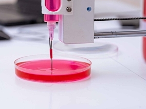
Now, researchers at Brigham and Women’s Hospital have developed a stretchable double-network hydrogel bioink system for microfluidic bioprinting of small-diameter vascular conduits. In Science Advances, they say the conduits recapitulate the structural and biological functions of native blood vessels.
The authors are Y. Shrike Zhang, PhD, principal of the Zhang Lab in the Division of Engineering in Medicine, C. Keith Ozaki, MD, director of vascular surgery research in the Department of Surgery and the Heart and Vascular Center, and colleagues.
Generation of Conduits
The bioink prepolymer is composed of sodium alginate and gelatin. It was crosslinked by calcium chloride and microbial transglutaminase, respectively, generating a double-networked hydrogel with excellent mechanical properties and biocompatibility.
Using microfluidic bioprinting, the researchers relied on two distinct approaches to engineer venous and arterial vessels:
- Venous conduits were generated by bioprinting monolayered hydrogel tubes, then seeding human umbilical vein endothelial cells in the lumens and human umbilical vein smooth muscle cells on the outer surfaces
- Arterial conduits need thicker muscular walls, so they were generated by bioprinting two layers: an outer layer encapsulated with human umbilical artery smooth muscle cells and an inner hydrogel layer; human umbilical artery endothelial cells were then seeded in the lumen of the inner wall
Results
Key observations were that:
- Both the venous and arterial conduits exhibited key properties of blood vessels, including high stretchability, apparent perfusability, selective permeability, and barrier performance
- The thicker smooth muscle layer of arterial conduits allowed them to constrict in response to a vasoconstrictor and dilate in response to a vasodilator
- The conduits proved feasible as an in vitro model of blood vessel infection with pseudotyped SARS-CoV-2 viral particles and the effects of antiviral drugs; they are expected to aid the study of other diseases and drugs
- Ex vivo anastomoses were successful when the conduits were connected to a mouse aorta or human popliteal vein, and in vivo anastomosis was accomplished with a mouse vena cava
The latter proof-of-concept test is direct evidence of the potential for in vivo vascular reconstructions with the new material, although further optimization is needed.