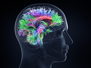
In 2020, the FDA announced the first clearance of a transcranial magnetic stimulation (TMS) system for adjunctive use in treating nicotine addiction. The device is designed to stimulate multiple brain regions because there is no consensus on optimal targets.
It’s now possible to link lesions in different brain locations to a common neuroanatomical substrate using the human connectome, a map of human brain connectivity. Using this approach, Michael D. Fox, MD, PhD, director of the Center for Brain Circuit Therapeutics at Brigham and Women’s Hospital, Juho Joutsa, MD, PhD, of the University of Turku, and colleagues have identified specific testable targets for therapeutic neuromodulation of nicotine addiction. The authors detail the results in Nature Medicine.
Voxel-wise Lesion–symptom Mapping
The researchers obtained CT/MRI data on 129 patients who were active daily smokers at the time of an acquired brain lesion. Afterward, 34 of them (26%) experienced nicotine addiction remission—they immediately quit smoking without difficulty, did not relapse, and reported no cravings. The others continued smoking.
Standard voxel-wise lesion–symptom mapping (VLSM) did not identify any lesion locations significantly correlated with addiction remission.
Lesion Network Mapping
Next, the team investigated functional connectivity between each lesion location and all other brain voxels. This lesion network mapping showed that although lesions associated with addiction remission occurred in many different brain locations, they were characterized by a specific pattern of brain connectivity:
- Positive connectivity to the dorsal cingulate, lateral prefrontal cortex, and insula
- Negative connectivity to the medial prefrontal and temporal cortex
In theory, the positive nodes in this addiction remission network represent ideal locations to place a focal lesion to disrupt addiction. Negative nodes should be ideal locations for excitatory brain stimulation, such as high-frequency TMS.
The connectivity pattern was reproduced when computed separately using two independent connectomes: one derived from 126 current smokers and another derived from 1,000 healthy volunteers.
Generalizability
In 186 patients who completed an alcoholism risk assessment, the connectivity profile of lesions associated with lower alcoholism risk was similar to the connectivity profile of lesions that disrupted nicotine addiction, even after adjustment for smoking status. Repeating the analysis using VLSM did not demonstrate similarity, so the concordance between the two sets of lesions was driven by network connectivity, not lesion location alone.
The concordance also proved to be specific to addiction risk. Maps generated for 10 other Minnesota Multiphasic Personality Inventory variables failed to match as well as the addiction risk map did, and the same was true of 27 maps generated using other neuropsychological variables.
Alignment With Previously Identified Targets
The brain locations that best matched the addiction remission map aligned with targets that have previously shown promise in treating addiction:
- The peak positive nodes were in the left frontal opercular cortex, which is adjacent to the left insula, and in the paracingulate gyrus, which is adjacent to the anterior cingulate
- The peak negative node was in the medial fronto-polar cortex, which overlaps the maximal electric field intensity of the TMS device recently cleared by the FDA for smoking cessation
Future Directions
The key takeaway from this study is that a network of brain regions, not just a single region, are involved in addiction remission—some positively and some negatively. Neuromodulatory treatments that excite and inhibit different regions simultaneously, such as multifocal electrode arrays, may prove useful.
