Management of lower gastrointestinal (GI) bleeding has become increasingly nuanced because of the aging of the population and the more frequent use of antiplatelet and anticoagulant medications.
Daniel J. Stein, MD, MPH, a gastroenterologist in the Department of Gastroenterology, Hepatology and Endoscopy at Brigham and Women’s Hospital, Joseph D. Feuerstein, MD, a gastroenterologist at Beth Israel Deaconess Medical Center, and a colleague recently reviewed for hospitalists the first-line diagnostic and treatment options when acute lower GI bleeding is suspected. Their paper in the Journal of Hospital Medicine includes a diagnostic algorithm that reflects American College of Gastroenterology and American College of Radiology (ACR) recommendations, guided by bleeding acuity and suspected diagnosis.
Endoscopy
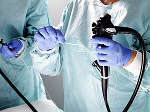
Several clinical presentations favor initial endoscopic evaluation. If rapid bowel preparation is possible, the best choice is often colonoscopy since it allows diagnosis and treatment in a single procedure. The optimal timing depends on hemodynamic stability, the quantity of blood loss, patient frailty, and suspected bleeding source.
Suspected neoplasm—Colonoscopy is the first choice
Suspected diverticular bleeding—In cases of massive hemorrhage, a colonoscopy may not be timely enough, but it’s indicated for slower bleeds because it can localize the site of bleeding and provide definitive hemostasis via endoscopic clipping or electrocautery
Colonic angioectasia and arteriovenous malformations—Managed similarly to suspected diverticular bleeding, although therapy is usually with argon plasma coaptation or clipping
Post-polypectomy hemorrhage—Should almost always be managed with sigmoidoscopy or full colonoscopy, depending on the location of the polypectomy; most cases can be treated with clipping or electrocautery
Upper GI (particularly rapid duodenal) bleeding—This may present with bright red blood per rectum; this requires urgent esophagogastroduodenoscopy as a first step
CT Angiography (CTA)
CTA obviates the need for bowel preparation and is most useful in cases of brisk hemorrhage, especially suspected diverticular bleeding. A study of prospectively collected data, published in JAMA Surgery, showed that including CTA before conventional angiography improves the identification of the bleeding source without worsening renal function.
Because CTA localizes the bleeding area, therapeutic angiography can be more selective. Therapeutic angiography without initial CTA is typically reserved for unstable patients (>5 units of blood in 24 hours or hemodynamic instability).
For patients with undifferentiated hemorrhagic shock and hematochezia, CTA can be the first localizing step as long as it’s rapidly available.
Tagged Red Cell Scans
The ACR guidelines categorize 99mTc-labeled red blood cell scintigraphy as “usually appropriate” for hemodynamically stable patients and “usually not appropriate” for hemodynamically unstable patients. CTA can generally be completed in a fraction of the time.
In a study by Dr. Feuerstein and colleagues, published in the American Journal of Roentgenology, the site of bleeding was accurately localized on 53% of CTA scans but only 30% of scintigraphic scans (P=0.008). Thus, physicians should interpret the results of tagged red cell scans as either positive or negative and not attempt to infer the location of the bleed.
Tagged red cell scanning may be considered for patients with significant renal impairment precluding the use of CTA. When positive within 2 hours, patients should undergo therapeutic angiography.
Surgery
Surgical evaluation is indicated for:
- Any suspected bleeding associated with severe ischemic colitis (prompt intervention needed)
- Ongoing, recurrent bleeding due to colorectal neoplasm
- Persistent bleeding associated with intestinal perforation or obstruction, peritonitis, intussusception, or recurrent massive GI bleed
- Uncontrolled diverticular bleeding that has failed first and second‐line therapies (rare)
With the help of the proposed algorithm, hospitalists can work with gastroenterologists and radiologists to create the most individualized treatment plan possible for each patient.
