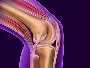
Tibial tuberosity osteotomy (TTO) is a common surgery for patellar instability and/or patellofemoral chondrosis. In most cases of anteromedialization TTO (the currently favored technique) patellar and trochlear lesions are easily accessible and the distal cortical hinge of the tubercle can be left intact. Such procedures can be abbreviated TTO-HI.
When patients have multiple defects in several compartments, though, the hinge of the tubercle is frequently detached (TTO-HD) and the tubercle fragment is flipped proximally. This fully exposes the knee joint and the articular surface of the patella, but Hoffa’s fat pad is more likely to dry out in room air than during TTO-HI.
Gergo Merkely, MD, of the Cartilage Repair Center in the Division of Sports Medicine at Brigham and Women’s Hospital, and colleagues wondered whether the extensive trauma and greater traction on the patella tendon might induce an inflammatory response that increases the risk of postoperative stiffness. In Cartilage, they report evidence that, indeed, TTO-HD is an independent risk factor for postoperative arthrofibrosis.
Methods
The research team reviewed data on 127 patients who had anteromedialization TTO performed between January 2007 and May 2017 by a single surgeon. All had a concomitant cartilage repair procedure, either osteochondral allograft transplantation or autologous chondrocyte implantation, and had at least two years of follow-up.
95 of the patients (59%) underwent TTO-HD, and 66 underwent TTO-HI (details of each procedure are given in the article). Postoperative rehabilitation for the two procedures was similar. The mean age at the time of surgery was 33, and the mean follow-up period was 4.6 years.
Results
In the TTO-HD group, significantly larger cartilage defects were repaired (mean 8.8 cm2 vs. 4.9 cm2 with TTO-HI; P<0.05), and the mean number of defects was greater (2 vs. 1.5; P<0.05).
The comparison of postoperative outcomes was:
- Need for subsequent surgery—60% of the TTO-HD group vs. 34% of the TTO-HI group (P<0.05)
- Arthrofibrosis as the reason for subsequent surgery (knee flexion <75° and/or lack of extension ≥15° in patients who underwent surgery because of knee stiffness)—31% vs. 6% (P<0.05)
- Chondroplasty, loose body removal, meniscus surgery or graft failure as reasons for subsequent surgery—No significant differences between groups
On multivariant analysis that controlled for the number and size of defects, the TTO-HD group was almost seven times more likely to develop postoperative arthrofibrosis than the TTO-HI group (OR, 6.5; 95% CI, 1.7–24.2; P<0.05).
Advice for Surgeons
In patients with symptomatic cartilage lesions, the process of early osteoarthritis has already begun. Early osteoarthritis is itself a risk factor for arthrofibrosis after injury or surgery, so patients who undergo cartilage repair procedures, especially those with large lesions, are already at risk to develop postoperative adhesions.
Certain cases require wide visualization and access, and therefore, the use of TTO-HD is recommended. Yet treating surgeons should be aware of the associated risk of postoperative arthrofibrosis and reintervention. TTO-HD should be used only judiciously, and patients should be carefully monitored postoperatively for signs of reduced range of motion.
