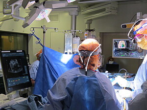
Intraoperative magnetic resonance imaging (iMRI) is an extremely useful tool in surgical brain tumor resection but also has significant drawbacks. For this reason, the Department of Neurosurgery at Brigham and Women’s Hospital is turning to an older—and perhaps surprising—technique to help guide tumor removal: ultrasound.
The Brigham has a long history of innovation in surgical care. In fact, notes Alexandra J. Golby, MD, director of image-guided neurosurgery, the first-ever use of iMRI took place at the hospital in the early 1990s. Today, Dr. Golby and her colleagues are leading the way in harnessing the potential of ultrasound to guide tumor resection.
“Ultrasound wasn’t a great technique 25 years ago,” she says. “That, along with the fact that it actually predates MRI, has meant some neurosurgeons don’t appreciate its utility using contemporary acquisition and analysis strategies. And so, we and others at the forefront of brain tumor surgery are creating a renaissance of the use of ultrasound.”
Advantages of Ultrasound Over iMRI
While acknowledging the many benefits of iMRI, Dr. Golby says, “One of our goals is to use lower-cost technology that is less resource-intensive, less disruptive to the surgical workflow and potentially more broadly available.”
To that end, the Brigham neurosurgery team has been relying more and more on ultrasound during brain tumor resection. Using ultrasound for this purpose dates back to the early 1980s (as published in Radiology). The technique subsequently fell from favor, due in large part to relatively poor image quality.
Over the past few decades, however, image quality has improved considerably. As a result, some companies are working to leverage ultrasound to guide neurologic, kidney, liver and other interventions. These efforts center on repackaging the technology to make it easier for surgeons to use themselves.
“That’s really appealing to surgeons because we do things with our hands, and we like having control,” Dr. Golby says. “New surgical ultrasound strategies allow us to move on from what we used to do—calling a radiologist into the OR to perform an ultrasound when we faced a difficult intraoperative challenge—to ultrasound being a device that we use regularly and know how to get the images we want without disrupting the operative workflow.”
Encouraging Ultrasound Adoption by Neurosurgeons
Compared with CT and MRI scans, ultrasound adoption among neurosurgeons is low. Dr. Golby believes this is partially attributable to habit: Neurosurgeons are simply more accustomed to using these other technologies.
Dr. Golby and her colleagues are collaborating with experts in computational image processing to enhance the images captured via ultrasound and make them more informative for surgeons. For instance, CT and MRI allow surgeons to view the brain in three cardinal planes—axial, coronal and sagittal. With ultrasound, in contrast, you create a view that is dependent on the exact position of the craniotomy and angle of the ultrasound probe.
“With some of these processing tools, it’s possible to do two things that are helpful,” she says. “One is to register the image to the preoperative cross-sectional images so that we can actually make correspondences between the image and, for example, an MRI. Second, we can essentially reconstruct the ultrasound. That involves sweeping the probe over the area of interest and reconstructing that image into a 3D dataset, which we can then explore using a pointer from the navigation system.”
Thanks to the advances taking place at the Brigham and elsewhere, Dr. Golby is confident that growing numbers of neurosurgeons will turn to ultrasound to guide procedures in the future.
“I think there’s gaining appreciation that by bringing contemporary ultrasound systems into the operating room and marrying them with novel image analysis and display strategies, it can be a very useful tool for surgeons,” she concludes.
