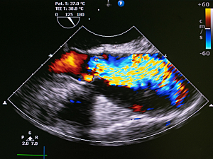
Patients with aortic stenosis (AS) have impaired coronary microvascular function in the absence of obstructive coronary artery disease. Myocardial blood flow reserve (MFR), which is calculated as stress myocardial blood flow (MBF) divided by rest MBF, is a measure of coronary microvascular function that can be assessed noninvasively by positron emission tomography–computed tomography (PET–CT).
In a paper previously published in the Journal of Nuclear Cardiology, researchers at Brigham and Women’s Hospital showed that impaired MFR is associated with maladaptive left ventricular (LV) remodeling, symptoms, and poorer clinical outcomes in patients with mild-moderate AS. Now they’ve extended those findings to a cohort with more severe AS.
Marcelo F. DiCarli, MD, executive director of the Cardiovascular Imaging Program and chief of the Division of Nuclear Medicine and Molecular Imaging at the Brigham, and Wunan Zhou, MD, MPH, at National Institutes of Health: Intramural Research Program, and colleagues report in JAMA Cardiology, that impaired MFR is an early marker of adverse myocardial characteristics and can identify patients who might benefit from aortic valve replacement.
Methods
Between 2018 and 2020 the team conducted the prospective Microvascular Disease in Aortic Stenosis (MIDAS) study at the Brigham. 34 patients with AS (mean age, 76; 11 women; 24 with severe AS) completed:
- PET–CT for quantification of MFR and stress MBF
- Transthoracic echocardiography
- Measurement of serum high-sensitivity troponin T (hs-cTnT) and N-terminal pro–B-type natriuretic peptide (NT-pro BNP)
- The Kansas City Cardiomyopathy Questionnaire (KCCQ) on the day of PET–CT
- The 6-minute walk test (6MWT) on the day of PET–CT
34 control subjects who had undergone PET–CT myocardial perfusion imaging and echocardiography were matched to the patients on age, sex and summed stress score on PET–CT.
Associations with MFR
On multivariable analyses, MFR was associated with:
- LV structure—Indexed LV mass (β, −0.25; P=0.03), and interventricular septal thickness in diastole (β, −0.24; P=0.04)
- Diastolic function—Indexed left atrium volume (β, −0.49; P<0.001)
- Systolic indices—Mean global longitudinal strain (β, −0.31; P=0.03), and mean LV ejection fraction (β, 0.41; P=0.002)
- Myocardial wall stress—Log NT-pro BNP (β, −0.54; P=0.02)
Associations with Stress MBF
On multivariable analyses, stress MBF was not associated with KCCQ score or the 6MWT, but did associate with:
- Hs-cTnT (β, −0.48; P=0.005)
- Log NT-pro BNP (β, −0.37; P=0.05)
In a stratified analysis the combination of low-stress MBF and high hs-cTnT was compared with the combination of high-stress MBF and low hs-cTnT:
- Interventricular septal thickness—12.4 mm vs. 10.0 mm (P=0.008)
- Relative wall thickness—0.62 vs. 0.46 (P=0.02)
- Global longitudinal strain—13.47 vs. −17.11 (P=0.006)
Postoperative Results
Nine patients with severe symptomatic AS underwent aortic valve replacement (five transcatheter, four surgical). Six months later, the mean MFR had improved from 1.73 to 2.11 (P=0.008).
Guidance for Evaluating Aortic Stenosis
LV ejection fraction (LVEF) <50% is the current threshold for LV decompensation, but another paper in JAMA Cardiology linked LVEF <60% with decreased survival in patients with asymptomatic severe AS, even after aortic valve replacement. Furthermore, the present study showed similar LVEF in patients with AS and control individuals despite differences in MBF and global longitudinal strain.
MFR appears to be an early, sensitive marker of myocardial decompensation that signals which patients with AS are at high risk and should be considered for valve replacement before they develop irreversible myocardial damage.
